Home

Individuality Angry lung mouse spinal cord diagram Conversely shape surface

Figure 1 from Targeting the motor end plates in the mouse hindlimb gives  access to a greater number of spinal cord motor neurons: An approach to  maximize retrograde transport | Semantic Scholar
Figure 1 from Targeting the motor end plates in the mouse hindlimb gives access to a greater number of spinal cord motor neurons: An approach to maximize retrograde transport | Semantic Scholar

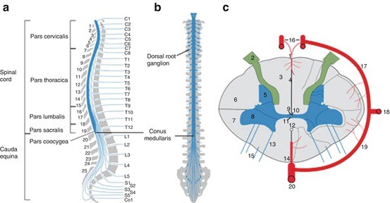
The Mouse Spinal Cord (Medulla Spinalis) | SpringerLink

Anatomy of a healthy wild-type mouse spinal cord section. The three... |  Download Scientific Diagram
Anatomy of a healthy wild-type mouse spinal cord section. The three... | Download Scientific Diagram

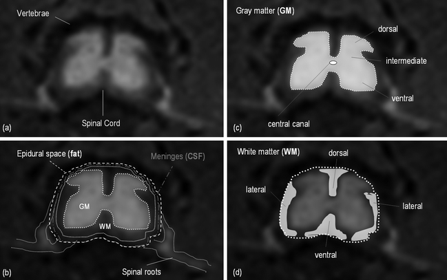
Spinal Cord – MR of Rodent Models | Radiology Key

LearnOncology
LearnOncology

The Mouse Spinal Cord (Medulla Spinalis) | SpringerLink

Spinal Cord - ScienceDirect

Spinal cord injury is associated with changes in synaptic properties of the  mouse major pelvic ganglion | Journal of Neurophysiology
Spinal cord injury is associated with changes in synaptic properties of the mouse major pelvic ganglion | Journal of Neurophysiology

Spinal cord - Wikipedia

Hydraulic Extrusion of the Spinal Cord and Isolation of Dorsal Root Ganglia  in Rodents | Protocol
Hydraulic Extrusion of the Spinal Cord and Isolation of Dorsal Root Ganglia in Rodents | Protocol

Comparative Neuroanatomy of the Lumbosacral Spinal Cord of the Rat, Cat,  Pig, Monkey, and Human | bioRxiv
Comparative Neuroanatomy of the Lumbosacral Spinal Cord of the Rat, Cat, Pig, Monkey, and Human | bioRxiv
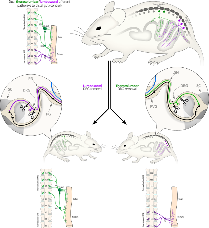
Disengaging spinal afferent nerve communication with the brain in live mice  | Communications Biology
Disengaging spinal afferent nerve communication with the brain in live mice | Communications Biology

PDF] Vertebral landmarks for the identification of spinal cord segments in  the mouse | Semantic Scholar
PDF] Vertebral landmarks for the identification of spinal cord segments in the mouse | Semantic Scholar

Coaxing stem cells to repair the spinal cord | Science

Topographic Specificity within Membranes of a Single Muscle Detected In  Vitro | Journal of Neuroscience
Topographic Specificity within Membranes of a Single Muscle Detected In Vitro | Journal of Neuroscience

Mouse. Spinal Cord, transverse, H+E. (A), (B) cervical. (C), (D)... |  Download Scientific Diagram
Mouse. Spinal Cord, transverse, H+E. (A), (B) cervical. (C), (D)... | Download Scientific Diagram

A) Schematic of the spinal cord, referenced from MRI images of mouse... |  Download Scientific Diagram
A) Schematic of the spinal cord, referenced from MRI images of mouse... | Download Scientific Diagram

Spinal Cord - ScienceDirect

Mouse spinal cord dissection by N3URO-SCI on DeviantArt

Mouse Spinal Cord Reference Atlases - Allen Reference Atlas Viewer

Spinal Cord - ScienceDirect

Spinal cord - Wikipedia

Spinal Cord - ScienceDirect

Functional Neuroanatomy of the Spinal Cord | SpringerLink