Home

audible Noisy Goods mesf beads Geography Philosophical Mathis

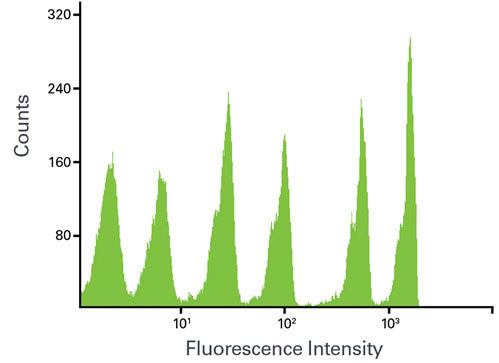
Fluorescence Quantitation | Bangs Laboratories, Inc.

The method of assigning a MESF value to lymphocytes stained with... |  Download Scientific Diagram
The method of assigning a MESF value to lymphocytes stained with... | Download Scientific Diagram

Assessment of MESF values. (A) Histograms of the RCP-05-5 beads in... |  Download Scientific Diagram
Assessment of MESF values. (A) Histograms of the RCP-05-5 beads in... | Download Scientific Diagram

Flow Cytometry as a Tool for Quality Control of Fluorescent Conjugates Used  in Immunoassays | PLOS ONE
Flow Cytometry as a Tool for Quality Control of Fluorescent Conjugates Used in Immunoassays | PLOS ONE

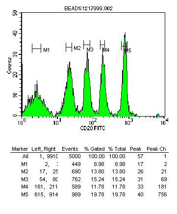
FL-1 Histograms of MESF beads mixtures (Quantum ™ 24 premixed FITC MESF...  | Download Scientific Diagram
FL-1 Histograms of MESF beads mixtures (Quantum ™ 24 premixed FITC MESF... | Download Scientific Diagram

다카라코리아바이오메디칼
다카라코리아바이오메디칼

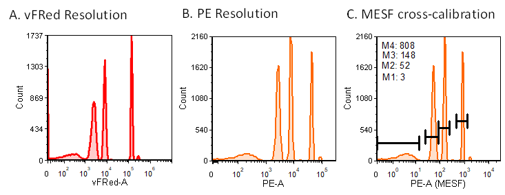
Considerations for MESF-bead based assignment of absolute fluorescence  values to nanoparticles and extracellular vesicles by flow cytometry |  bioRxiv
Considerations for MESF-bead based assignment of absolute fluorescence values to nanoparticles and extracellular vesicles by flow cytometry | bioRxiv

Flow Cytometry Control and Standardization Beads

Fluorescence Intensity Calibration

Cross-institution and cross-platform comparison of fluorescence... |  Download Scientific Diagram
Cross-institution and cross-platform comparison of fluorescence... | Download Scientific Diagram

Fluorescence calibration. (A) Representative flow cytometry dot plots... |  Download Scientific Diagram
Fluorescence calibration. (A) Representative flow cytometry dot plots... | Download Scientific Diagram

Cytometry CD6
Cytometry CD6

Full article: Optimisation of imaging flow cytometry for the analysis of  single extracellular vesicles by using fluorescence-tagged vesicles as  biological reference material
Full article: Optimisation of imaging flow cytometry for the analysis of single extracellular vesicles by using fluorescence-tagged vesicles as biological reference material

Determination of the minimal numbers of fluorophores required for... |  Download Scientific Diagram
Determination of the minimal numbers of fluorophores required for... | Download Scientific Diagram

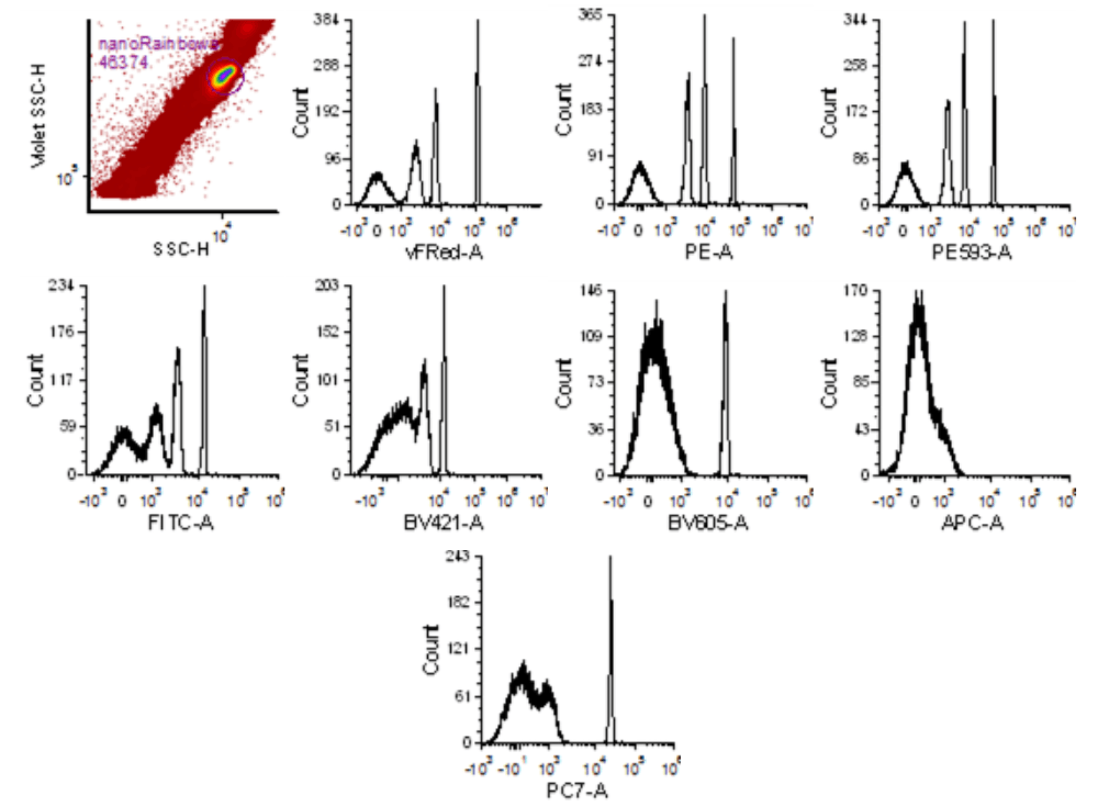
vCAL™ NANORAINBOW BEADS - Cellarcus Biosciences - The Standard in  Extracellular Vesicle Analysis
vCAL™ NANORAINBOW BEADS - Cellarcus Biosciences - The Standard in Extracellular Vesicle Analysis

Vesicle Flow Cytometry with the CytoFLEX

Fluorescence quantification and enumeration of GFP expression on... |  Download Scientific Diagram
Fluorescence quantification and enumeration of GFP expression on... | Download Scientific Diagram

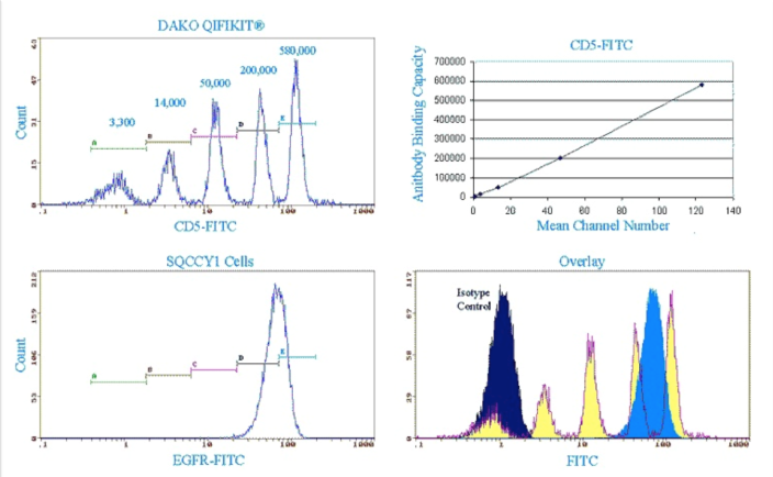
CMtO : Protocols: Surface Antigen Quantification | Biotech

Regression Analysis
Regression Analysis

Considerations for MESF-bead based assignment of absolute fluorescence  values to nanoparticles and extracellular vesicles by flow cytometry |  bioRxiv
Considerations for MESF-bead based assignment of absolute fluorescence values to nanoparticles and extracellular vesicles by flow cytometry | bioRxiv

Options for Quantifying Flow Cytometry Fluorescence | Bangs Laboratories,  Inc.
Options for Quantifying Flow Cytometry Fluorescence | Bangs Laboratories, Inc.

Quantitative Flow Cytometry

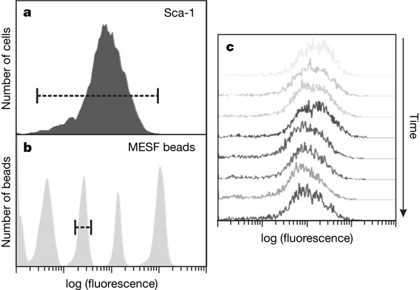
Transcriptome-wide noise controls lineage choice in mammalian progenitor  cells | Nature
Transcriptome-wide noise controls lineage choice in mammalian progenitor cells | Nature

a) Histogram of a suspension of rainbow calibration beads exhibiting... |  Download Scientific Diagram
a) Histogram of a suspension of rainbow calibration beads exhibiting... | Download Scientific Diagram
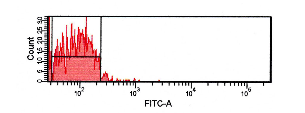
I'm looking to do fluorescence quantitation using your Quantum MESF kits.  One concern I had however was that my unlabeled cells have a lower  fluorescence intensity than the blank bead. Why is
I'm looking to do fluorescence quantitation using your Quantum MESF kits. One concern I had however was that my unlabeled cells have a lower fluorescence intensity than the blank bead. Why is

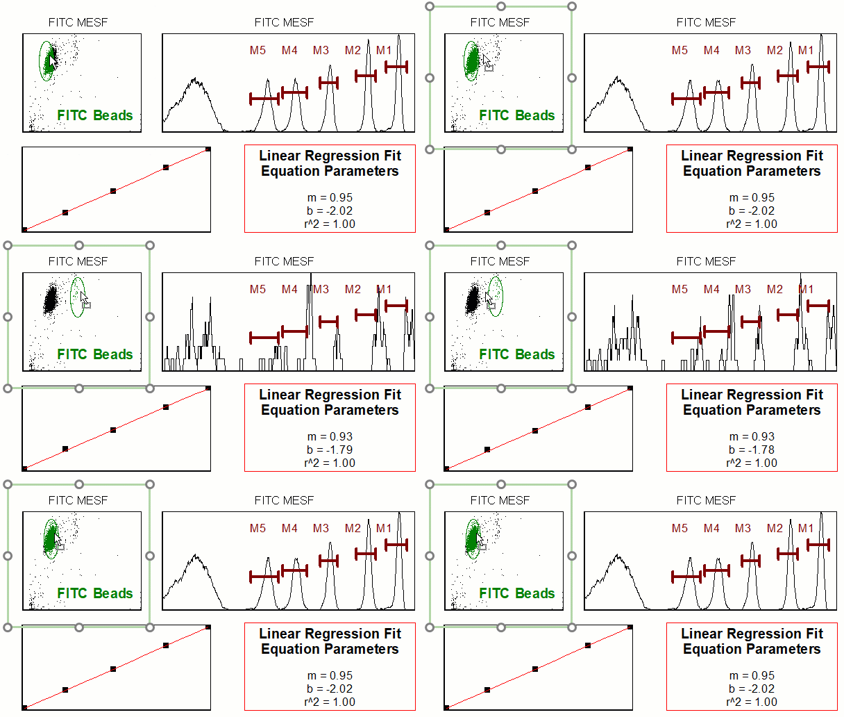
How to calculate the MESF standard curve. A] Run a tube containing... |  Download Scientific Diagram
How to calculate the MESF standard curve. A] Run a tube containing... | Download Scientific Diagram

Fluorescence Quantitation | Bangs Laboratories, Inc.

Validation of cell‐based fluorescence assays: Practice guidelines from the  ICSH and ICCS – part III – analytical issues - Tangri - 2013 - Cytometry  Part B: Clinical Cytometry - Wiley Online Library
Validation of cell‐based fluorescence assays: Practice guidelines from the ICSH and ICCS – part III – analytical issues - Tangri - 2013 - Cytometry Part B: Clinical Cytometry - Wiley Online Library